Research Overview

Background
Mitochondria are an ideal system to study the geometry of intracellular structures because their morphology ranges from small and discrete to large and very dynamic interconnected tubular networks. Mitochondria perform a vital role in respiration and cellular energy production, and mitochondrial function and morphology are implicated in blindness, neurological and respiratory diseases, cancer and aging. Budding yeast has long been a model organism of choice for studying the molecules involved in mitochondrial morphology and function as it can easily survive in the absence of respiration. The success of budding yeast as a genetic and systems biology model organism makes it a clear choice for my long-term goals.
In budding yeast, mitochondria compose a 3D network of membrane bound tubules localized at the cell periphery. The network undergoes constant remodeling due to balanced fusion and fission dynamics.
research Topics (click on topic for more information)
Mitochondrial Networks in budding yeast
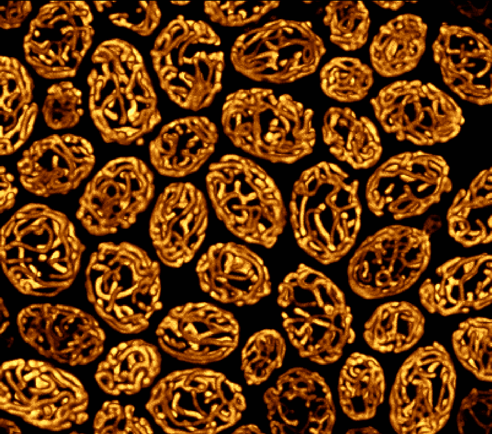
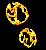
Structured Illumination Microscopy (SIM) of mitochondria in fixed yeast cells
3D-deconvolution microscopy of mitochondria in live yeast cells. 1 minute intervals for 10 minutes (movie plays 120x)
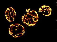
Top: Volume view of mitochondria in live yeast cells growing in glycerol
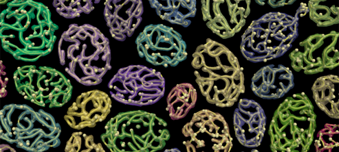
Bottom: Mitographs overlayed onto 3D reconstructions of mitochondria
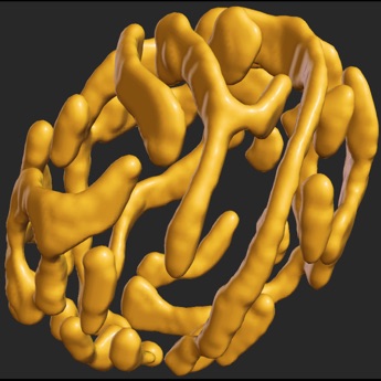
3D surface reconstruction of a mitochondrial network in an unbudded yeast cell. Click on image to explore the network in 3D.